Peri-Oral Ageing: The Clinical Picture, and How We Treat It
A patient education and clinical reference guide, Mapp Aesthetics
Contents
Overview
Anatomy of the Peri-Oral Region
The Pathophysiology of Peri-Oral Ageing
Contributing and Accelerating Factors
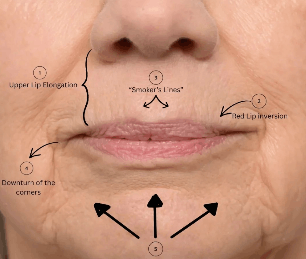
The Clinical Signs of Peri-Oral Ageing
Assessment and Classification
Treatment Option 1: Muscle-Relaxing Treatment
Treatment Option 2: Skin Boosters
Treatment Option 3: Structural and Volume Support
Treatment Option 4: Resurfacing and Energy-Based Devices
Treatment Option 5: Medical Skincare and Modifiable Risk Factors
Building a Combination Treatment Plan
Patient Selection, Contraindications and Cautions
Pre-Treatment Preparation
Aftercare
Complications and Safety
Realistic Expectations
Prevention and Long-Term Maintenance
Special Patient Populations
Frequently Asked Questions
Glossary
Important Information
1. Overview
The peri-oral region, meaning the skin, muscle, fat and bone surrounding the mouth, is among the earliest and most conspicuous areas of the face to display visible ageing. It is also one of the most functionally demanding regions of the body. The mouth is in near-constant motion during speech, eating, drinking and emotional expression, and the tissue surrounding it is structurally among the thinnest and least supported on the face.
The combination of high mechanical load and low structural reserve explains why change here appears early, progresses steadily, and is disproportionately noticeable to the patient. Vertical lip lines, thinning of the vermilion, downturning of the oral commissures and loss of definition at the lip border can shift the perceived age, mood and even health of the lower face, often well before a patient feels they have aged elsewhere.
Patients frequently describe these changes as "smoker's lines", a term that has persisted despite the majority of affected patients being lifelong non-smokers. It is a misleading label. The underlying process is multifactorial and largely universal. Smoking is an accelerant, not a prerequisite.
The purpose of this guide is to explain, in clinical detail, why peri-oral ageing occurs, how it is assessed, and what the evidence-based, non-surgical treatment options are, including their mechanisms, timelines, limitations and risks.
Peri Oral Treatment Area
2. Anatomy of the Peri-Oral Region
Effective treatment of this region depends entirely on an accurate understanding of its anatomy. The peri-oral area is anatomically dense, highly vascular and functionally critical.
2.1 Aesthetic boundaries and subunits
The peri-oral region is conventionally bounded:
Superiorly by the base of the nose (subnasale) and the nasolabial folds
Laterally by the nasolabial folds extending inferiorly toward the labiomandibular folds
Inferiorly by the labiomental crease and the chin
Within this region, the recognised aesthetic subunits are the two lateral upper lip subunits, the central philtral subunit, the lower lip and the chin. Treatments and any resurfacing are ideally planned by subunit to avoid visible demarcation lines.
Key surface landmarks include:
Vermilion: the reddish, keratinised but non-hair-bearing mucocutaneous portion of the lip
Vermilion border, or "white roll": the slightly raised ridge of pale skin at the junction between vermilion and cutaneous lip. This is a critical light-reflecting structure that blurs with age.
Cupid's bow: the double-arched contour of the upper vermilion border
Philtral columns: the paired vertical ridges running from the nasal base to Cupid's bow, formed by decussating fibres of the orbicularis oris
Oral commissures: the corners of the mouth
Labiomental crease: the horizontal fold between the lower lip and chin
2.2 The skin
Peri-oral skin has several distinguishing features that predispose it to early ageing:
A thin dermis with a relatively low collagen density compared with cheek skin
Minimal subcutaneous fat, particularly over the cutaneous upper lip, meaning the dermis sits almost directly on muscle
A high density of pilosebaceous units in the upper lip, which contributes to a fine but irregular surface texture
Direct dermal attachment to the underlying orbicularis oris muscle via short, dense fibrous septae. This is a defining anatomical feature in peri-oral rhytid formation, because it means every muscle contraction is transmitted directly and efficiently into the overlying skin.
Little to no protective bony convexity, unlike the forehead or malar region
2.3 The musculature
The peri-oral musculature is a complex, interdigitating sphincteric and radial system.
The sphincter
Orbicularis oris is a sphincteric muscle organised into two functional components: the pars marginalis (deep, adjacent to the vermilion, responsible for lip pursing and eversion) and the pars peripheralis (superficial and peripheral, contributing to lip closure and compression). Its fibres run predominantly circumferentially, meaning horizontally around the mouth. Because dermal rhytides form perpendicular to the direction of underlying muscle fibre contraction, this circumferential orientation is precisely why peri-oral lines are vertical.
The elevators of the upper lip
Levator labii superioris
Levator labii superioris alaeque nasi
Levator anguli oris
Zygomaticus major and minor
Risorius
The depressors of the lower lip and commissure
Depressor anguli oris (DAO), the principal driver of downturned mouth corners
Depressor labii inferioris
Mentalis, responsible for chin dimpling and lower lip protrusion
Platysma, whose superior fibres contribute to lower face and commissure depression
The modiolus
A dense, mobile fibromuscular node located approximately 1 cm lateral to each oral commissure, where the orbicularis oris, DAO, levator anguli oris, zygomaticus major, risorius, buccinator and platysma converge. The modiolus is the pivot point of lower facial expression. Its position descends with age, and it is an anatomically hazardous zone. Treatment placed carelessly here can cause profound functional impairment of the mouth.
2.4 Vascular anatomy
The peri-oral region is one of the most richly vascularised areas of the face, and this is the primary source of procedural risk.
The facial artery ascends from the inferior mandibular border, anterior to the masseter, and gives rise to the inferior labial artery and the superior labial artery, typically near the oral commissure.
The labial arteries most commonly run submucosally or intramuscularly, deep to the orbicularis oris and posterior to the vermilion. Their course is highly variable between individuals and even between sides in the same individual. Studies report submucosal positioning in the majority of cases, with intramuscular and subcutaneous variants also documented.
The superior labial artery gives off the septal and alar branches supplying the columella and nasal base.
Critically, the facial artery system anastomoses with the ophthalmic artery via the angular and dorsal nasal arteries. This creates a potential retrograde embolic pathway from the peri-oral region to the retinal circulation, and it is the anatomical basis for the rare but catastrophic complication of blindness following facial treatment.
Venous drainage occurs via the facial vein into the internal jugular system, with anastomotic connections to the pterygoid plexus and cavernous sinus.
2.5 Innervation
Sensory, via the trigeminal nerve (CN V)
Infraorbital nerve (V2) exits the infraorbital foramen approximately 5 to 8 mm below the infraorbital rim, supplying the upper lip, lateral nose and lower eyelid
Mental nerve (V3) exits the mental foramen, typically between the first and second premolar roots, supplying the lower lip and chin
Both are relevant to dental block anaesthesia and to the localised sensory disturbance occasionally reported after treatment.
Motor, via the facial nerve (CN VII)
Buccal branches supply the orbicularis oris, buccinator and upper lip elevators
Marginal mandibular branch supplies the DAO, depressor labii inferioris and mentalis. It runs along or below the mandibular border and is vulnerable in the lateral lower face.
2.6 The skeletal and dental foundation
The soft tissues of the peri-oral region are entirely dependent on their bony and dental scaffold.
Maxilla and pyriform aperture: the pyriform aperture widens and its angle becomes more obtuse with age, withdrawing support from the nasal base and central upper lip
Maxillary alveolar ridge: resorbs, particularly with tooth loss, reducing anterior projection of the upper lip
Mandible: undergoes reduction in both height and angle, with the mandibular angle becoming more obtuse
Mental foramen: with alveolar resorption the foramen effectively migrates superiorly toward the alveolar crest, an important consideration in chin and pre-jowl treatment
Dentition: attrition, loss or ill-fitting prostheses collapse vertical dimension and directly worsen peri-oral folding and commissure downturn
A patient with significant dental compromise will have a structurally different, and treatment-limited, peri-oral region compared with an age-matched patient with intact dentition.
2.7 Fat compartments
The peri-oral region contains discrete, septated fat compartments rather than a continuous fat layer:
Superior and inferior labial (orbicularis oris) fat compartments
Superficial and deep nasolabial fat
The deep medial cheek fat, whose deflation contributes to nasolabial deepening
Buccal fat, whose descent contributes to jowling and secondary commissure heaviness
Deep chin fat and the mentalis fat pad
These compartments do not deflate uniformly. Selective deflation and inferior displacement, rather than global atrophy, is the pattern most commonly observed.
3. The Pathophysiology of Peri-Oral Ageing
Peri-oral ageing is better understood as simultaneous change occurring across all five anatomical layers, being skin, superficial fat, muscle, deep fat and bone, rather than as a skin problem.
Fine lines above the lips
3.1 Skin and dermis
Intrinsic, or chronological, ageing
Dermal collagen content declines by an estimated 1% per year from approximately the third decade onward, with accelerated loss in the perimenopausal and early postmenopausal period.
The ratio of type III to type I collagen shifts, and the remaining collagen becomes increasingly cross-linked, disorganised and mechanically stiff.
Elastin fibres fragment and degrade, and functional elastogenesis in adult skin is minimal, meaning elastin loss is largely irreversible.
Glycosaminoglycans, the water-binding molecules of the dermis, decline substantially, reducing the dermis's water-binding capacity and producing the characteristic dehydrated, crepey appearance.
The dermal to epidermal junction flattens as rete ridges efface, reducing the surface area for nutrient exchange and weakening the mechanical bond between epidermis and dermis. This flattening is a major contributor to the fine, crinkled surface texture of the ageing upper lip.
Fibroblast number and secretory activity decline, and remaining fibroblasts become senescent, adopting a pro-inflammatory secretory phenotype that further degrades the matrix.
Epidermal turnover slows and the stratum corneum barrier becomes less efficient, increasing transepidermal water loss.
Extrinsic, or photo-, ageing
UVA radiation penetrates to the reticular dermis and generates reactive oxygen species, upregulating matrix metalloproteinases (MMP-1, MMP-3, MMP-9) which enzymatically degrade collagen and elastin.
UV simultaneously downregulates procollagen synthesis via suppression of TGF-beta signalling, a dual insult of accelerated breakdown and reduced production.
Chronic exposure produces solar elastosis, the accumulation of amorphous, dysfunctional elastotic material in the dermis, histologically characteristic of photoaged skin.
The cutaneous upper lip is horizontally oriented and highly sun-exposed, receiving a UV dose comparable to the nose and forehead, while being far less frequently protected. Sunscreen application typically stops at the lip margin.
3.2 Muscle
The orbicularis oris is in near-continuous use. Over decades, cumulative contraction produces repetitive dermal folding along fixed lines.
Once dermal collagen and elastin can no longer restore the skin to its resting configuration, a dynamic line becomes a static, etched rhytid.
Peri-oral muscles also demonstrate age-related hypertonicity and shortening. DAO hyperactivity in particular becomes more pronounced, actively pulling the commissures inferiorly.
Partial inversion of the vermilion occurs as the orbicularis contracts and shortens, reducing visible red lip show independently of true volume loss.
3.3 Fat
Deflation of the superficial and deep labial fat compartments reduces lip projection and eversion.
Deflation of the deep medial cheek fat and descent of the superficial nasolabial fat deepen the nasolabial fold and add weight above the commissure.
Inferior migration of the buccal fat and jowl formation places downward load on the modiolus and labiomandibular region, producing or worsening marionette lines.
The retaining ligaments, particularly the mandibular ligament, remain fixed while surrounding tissue descends, creating the tethering point that defines the pre-jowl sulcus and the medial extent of the jowl.
3.4 Bone
Bone loss is not passive shrinkage. It is regionally selective resorption.
Resorption at the pyriform aperture, maxilla and anterior alveolus withdraws the foundation beneath the upper lip, allowing it to lengthen, flatten and roll inward.
Mandibular changes reduce chin projection and alter the labiomental relationship.
Because skin surface area does not reduce in proportion to skeletal volume loss, the result is relative soft tissue excess over a contracted frame, one of the principal drivers of folding, redundancy and rhytid formation.
3.5 The final common pathway
Increased mechanical loading from muscle activity, applied to a structurally weakened dermis through collagen, elastin and glycosaminoglycan loss, over a contracting foundation of bone and fat volume loss, with impaired repair capacity from fibroblast senescence and MMP upregulation, produces the characteristic clinical picture of peri-oral ageing.
This is why single-modality treatment so often disappoints. Relaxing a muscle does not restore a dermis, and hydrating a dermis does not restore a skeleton.
Chronic sleep restriction, high glycaemic diet producing advanced glycation end-products, chronic inflammatory disease, chronic corticosteroid use
Prior surgery or trauma
Cleft repair, previous excisions or scarring alter muscle vectors and tissue compliance
5. The Clinical Signs of Peri-Oral Ageing
Clinically, patients present with some combination of the following.
Skin and surface changes
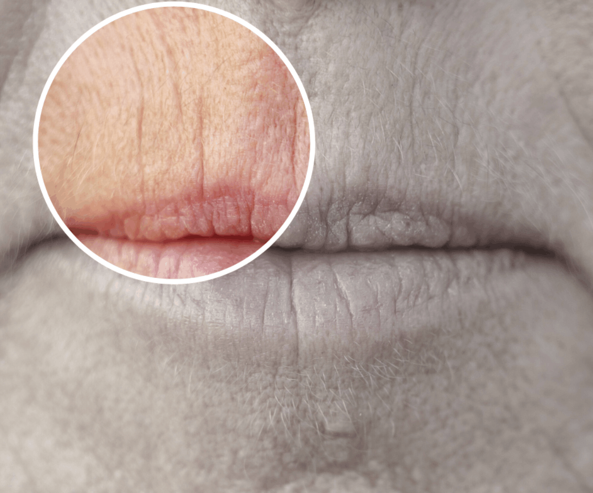
Vertical rhytides of the cutaneous upper lip, and less commonly the lower lip, which may be dynamic, static, or both
Crepey, finely wrinkled skin texture at rest
Loss of the light-reflecting definition of the vermilion border, causing lipstick migration or bleeding into fine lines
Dyspigmentation, actinic damage and telangiectasia
Actinic cheilitis of the lower lip in significantly sun-damaged patients, which requires assessment to exclude dysplasia
Structural and volumetric changes
Reduced vermilion show, particularly of the upper lip
Loss of Cupid's bow definition and flattening of the philtral columns
Lengthening of the cutaneous upper lip with reduced or absent maxillary incisor show at rest and on smiling
Inversion or inward rolling of the vermilion
Flattening of the lip's anterior projection in profile
Positional and expressive changes
Downturned oral commissures, giving a resting expression that reads as sad, tired or disapproving
Deepening of the labiomandibular, or marionette, folds
Pre-jowl sulcus formation and loss of a clean jawline
Deepening of the nasolabial folds
Peau d'orange chin dimpling from mentalis hyperactivity
Deepening or, alternatively, effacement of the labiomental crease
Functional changes in advanced cases
Oral commissure moisture pooling, leading to angular cheilitis
Difficulty with lipstick retention or with denture fit
6. Assessment and Classification
A structured assessment is the principal determinant of treatment outcome. The purpose is to establish which layer or layers are driving the presenting concern.
6.1 History
Presenting concern in the patient's own words, and what specifically bothers them
Duration and rate of progression
Smoking, vaping, sun exposure and photoprotection habits
Dental history, including recent extractions, dentures, orthodontics or planned dental work
Menopausal status and hormone therapy
Occupation and hobbies with peri-oral relevance, such as wind instruments, professional singing, voice work, scuba diving and swimming
Full medical history, medications (particularly anticoagulants, antiplatelets, immunosuppressants and aminoglycosides), and allergies
History of cold sores (herpes labialis)
Previous aesthetic treatment in the area, including type, timing and outcome
Psychological screening, including a low threshold for identifying body dysmorphic disorder
6.2 Examination
Assessment is performed at rest, in animation and in profile, under consistent lighting.
Examination area around the lips and lines under the nose
Vermilion height and upper to lower lip ratio. A commonly cited aesthetic reference is approximately 1 to 1.6.
Cutaneous upper lip length, measured subnasale to upper vermilion border. Commonly quoted normative ranges are approximately 15 to 18 mm in women and 18 to 22 mm in men, with increases indicating lip lengthening.
Maxillary incisor show at rest, typically 2 to 4 mm in younger patients and reducing with age
Oral commissure position relative to the intercommissural line
Symmetry, including pre-existing asymmetry, documented before treatment
Philtral column definition and Cupid's bow projection
In animation
Rhytid depth on maximal pursing, differentiating dynamic from static components
DAO activity on forced commissure depression
Mentalis activity and chin dimpling
Smile pattern and gingival show
Lip competence and speech clarity
In profile
Lip projection relative to standard reference lines
Chin projection and labiomental angle
Nasolabial angle
6.3 Classification tools
Standardised scales support consistent documentation and outcome measurement:
Perioral Lines at Rest and at Maximum Contraction scales, which are validated photonumeric gradings of peri-oral rhytid severity
Lemperle Wrinkle Assessment Scale, from 0 (none) to 5 (very deep), applicable to the upper lip
Fitzpatrick Wrinkle and Elastosis Scale, which grades both wrinkling and the degree of elastosis
Glogau Photoageing Classification, types I to IV, guiding the choice of resurfacing intensity
Fitzpatrick Skin Phototype I to VI, critical for predicting post-inflammatory hyperpigmentation risk with any resurfacing or energy-based modality
Validated lip fullness and oral commissure scales
6.4 Standardised photography
Baseline photography is mandatory. Standard views comprise frontal at rest, frontal in maximal pursing, frontal smiling, and bilateral obliques and profiles, with fixed lighting, distance, background and head position. Without standardised imaging, both patient and clinician consistently misjudge the degree of change achieved.
6.5 The diagnostic question
At the end of assessment, one question should be answerable:
Are these lines predominantly dynamic and movement-driven, static and skin-quality driven, structural and support-driven, or, most commonly, a combination, and in what proportion?
The answer dictates the treatment plan. Treating a static, elastotic rhytid with muscle relaxation alone will fail. Treating a purely dynamic line with a skin booster alone will underdeliver.
7. Treatment Option 1: Muscle-Relaxing Treatment
7.1 Rationale
Where vertical lip lines are driven predominantly by repetitive orbicularis oris contraction, reducing the strength of that contraction reduces the mechanical folding of the overlying skin. Over time, this also permits a degree of passive dermal recovery, as the skin is no longer being repeatedly creased along the same lines.
7.2 Mechanism of action
The treatment works at the neuromuscular junction. Following administration, the product is taken up by presynaptic cholinergic nerve terminals, where it interferes with the protein complex responsible for fusing acetylcholine-containing vesicles with the nerve terminal membrane. Without this fusion, acetylcholine cannot be released into the synaptic cleft, and the muscle fibre does not receive its contraction signal.
The effect is temporary and reversible. The blockade is not permanent because the nerve terminal responds by sprouting new axonal branches and, over subsequent weeks to months, the original terminal recovers function. Clinically, this manifests as a gradual return of movement.
Importantly, the effect is local and dose-dependent. The intent in the peri-oral region is partial weakening, never full paralysis, because full paralysis of the oral sphincter would be functionally unacceptable.
7.3 Treatment approach
Product is placed superficially and in small quantities along the cutaneous upper lip, close to the vermilion border, at a small number of symmetrically distributed points.
Doses used in this region are substantially lower than those used in the upper face, typically by an order of magnitude, because the therapeutic window is narrow.
Placement is deliberately kept away from the oral commissures and the modiolus to avoid affecting the muscles of the commissure complex.
Placement depth is kept intradermal or immediately subdermal rather than deep intramuscular, to limit diffusion into the pars marginalis.
Symmetry of dosing is essential. The mouth is the most scrutinised region of the face, and asymmetry here is immediately apparent.
Where DAO hyperactivity is contributing to commissure downturn, separate treatment of the DAO may be considered. This is a distinct treatment with distinct anatomical considerations and is planned independently.
Treatment takes a few minutes. Discomfort is mild and brief. No anaesthetic is routinely required, although topical anaesthetic can be used for needle-sensitive patients.
7.4 Suited to
Dynamic vertical lip lines, meaning lines that appear or deepen markedly on pursing, whistling or drinking through a straw
Patients in their thirties and forties using treatment preventatively, before lines become etched
Patients not yet ready for volume-based or resurfacing treatment
Use as a component of a combination plan, reducing ongoing mechanical stress while other modalities improve skin quality
7.5 Results timeline
Time pointExpected findingDay 0No visible changeDay 3 to 5Onset of softening becomes noticeableDay 10 to 14Peak effect, and the formal review pointWeek 4Stable clinical resultApproximately 4 to 8 weeksMovement begins to return in most patientsApproximately 8 to 12 weeksFull return of movement in some patients
Duration in the peri-oral region is consistently shorter than in the upper face, where three to four months is typical. This is expected and is not a treatment failure. The reasons are the low doses used, the very high baseline activity of the muscle, and its large motor unit turnover.
Review at two weeks after a first treatment is recommended so that dose and placement can be refined for subsequent sessions.
7.6 Downtime and recovery
Minimal. Small marks at each treatment point and occasional pinpoint bruising may be present for a short period and are readily concealed with makeup after 24 hours.
7.7 Possible side effects
Common and self-limiting
Small transient papules at each treatment point
Pinpoint bruising
Mild swelling
Brief tenderness
Headache
Functional, dose-related and temporary
Difficulty whistling, the most commonly reported functional effect
Difficulty drinking through a straw or from a narrow-rimmed vessel
Altered articulation of plosive consonants (p, b, m) and labiodental sounds (f, v), usually subtle, but significant for professional voice users
Difficulty with wind instruments, often a contraindication for professional players
Sensation of lip flatness, stiffness or reduced dexterity
Mild lip inversion or reduced vermilion show
Minor drooling or difficulty retaining liquid, in over-treatment
Uncommon
Asymmetry of the smile
Oral incompetence, typically resulting from excessive dose or inappropriately deep or lateral placement
Localised infection at a treatment point
Rare
Hypersensitivity reaction
Distant spread of effect with systemic symptoms. This is extremely rare at aesthetic doses, but patients should be advised to report any difficulty swallowing, difficulty breathing, or generalised weakness immediately.
7.8 The critical limitation
This treatment reduces movement. It does not improve skin quality.
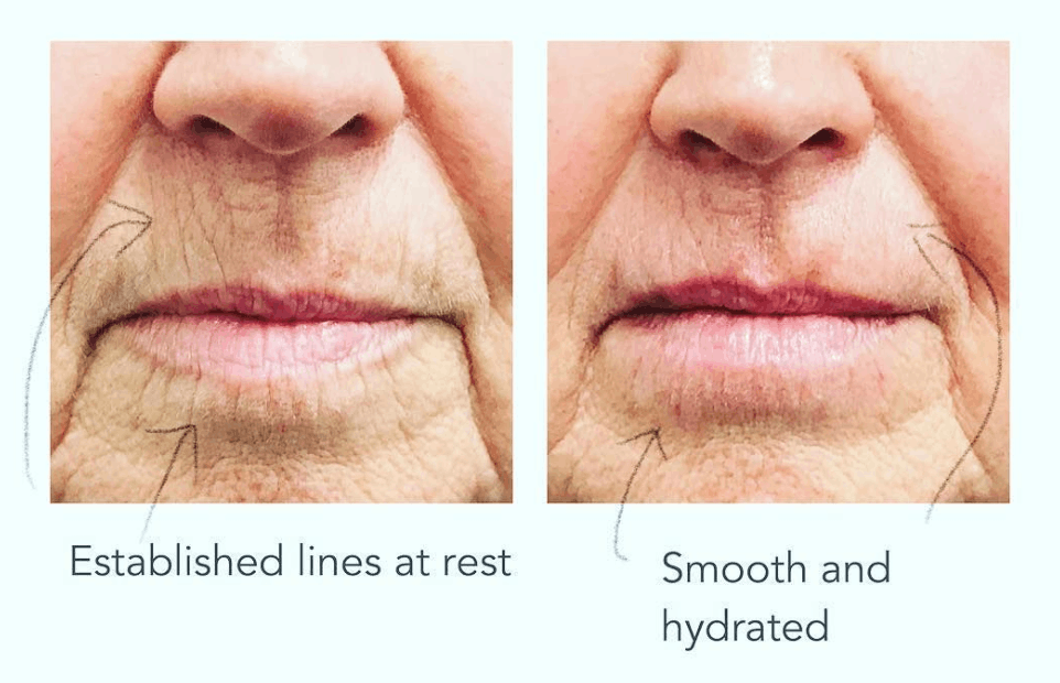
If the dermis is thin, dehydrated, elastotic and etched, softening muscle contraction will improve lines in animation while leaving them substantially unchanged at rest. Patients presenting primarily with static, resting lines who are treated with muscle relaxation alone are a frequent source of disappointment in this region.
This is not a shortcoming of the treatment. It is a mismatch between the treatment and the underlying pathology. It is also the clearest argument for combination therapy.
8. Treatment Option 2: Skin Boosters and Bio-Remodelling
Skin boosters remain one of the most under-utilised and under-appreciated treatments for peri-oral ageing. Where muscle-relaxing treatment addresses movement, skin boosters address the substrate, meaning the quality, hydration, thickness and elasticity of the dermis itself.
8.1 Rationale
The core deficits in peri-oral skin ageing are reduced dermal water content, reduced collagen and elastin, and a thinned, disorganised extracellular matrix. Skin boosters are designed to intervene directly at this level, rather than to soften a single line or restore a volumetric contour.
8.2 Mechanism of action
Product is placed into the dermis, not deep into the subcutaneous plane and not into muscle, where it acts through two complementary pathways.
1. Immediate hydration, a physical mechanism
The product is strongly hygroscopic, binding many times its own weight in water. This restores dermal water content, improving turgor, light reflection and surface smoothness. This effect is apparent early, often within one to two weeks, and accounts for the initial glow patients report.
2. Fibroblast stimulation, a biological mechanism
This is the more clinically significant and more durable effect. Restoring dermal hydration and volume mechanically stretches the resident fibroblasts. Fibroblasts are mechanosensitive cells, and physical stretch triggers mechanotransduction signalling that reactivates their synthetic programme.
Documented downstream effects include:
Increased synthesis of type I and type III collagen
Increased elastin production
Increased production of the skin's own water-binding molecules
Downregulation of matrix metalloproteinases, reducing ongoing matrix degradation
Improved dermal thickness and echogenicity on ultrasound assessment
Reduced markers of fibroblast senescence
In effect, the treatment does not simply add hydration. It restarts the skin's own repair machinery. This is why the clinical improvement continues to build for weeks after the product itself has been metabolised, and why results substantially outlast the residence time of the product.
8.3 Treatment approach
The area is cleansed and topical anaesthetic applied for approximately 20 to 30 minutes.
Product is delivered into the dermis via a series of small, precisely placed deposits, commonly using a papular or micro-bolus technique, sometimes with linear threading or fanning depending on the area and product characteristics.
Placement is planned across the cutaneous upper lip, the peri-oral field, and where indicated is extended to the chin, jawline and lower cheeks. Peri-oral skin does not exist in isolation, and treating an isolated square of skin creates a visible demarcation.
A fine needle or a cannula may be used depending on the technique and region.
Treatment typically takes 20 to 30 minutes including anaesthetic time.
8.4 Protocol
1 to 2 sessions, spaced approximately 4 weeks apart
Some protocols use a third session in significantly photodamaged or very thin skin
Results last approximately 9 to 12 months
Maintenance sessions are usually recommended at 6 to 12 month intervals depending on individual response, skin quality and lifestyle factors
8.5 Suited to
Static vertical lip lines, visible at rest and not only on animation
Thin, crepey, dehydrated peri-oral skin
Patients with fine surface etching rather than deep folds
Smokers and former smokers with established dermal damage
Peri- and post-menopausal patients with generalised dermal thinning
Patients seeking skin rejuvenation without volumetric change to the lips
Patients who explicitly do not want larger or altered-looking lips
Combination with muscle-relaxing treatment for mixed dynamic and static lines
8.6 Results timeline
Time pointExpected finding24 to 48 hoursSmall papules at each placement point settleWeek 1 to 2Visible improvement in hydration, light reflection and surface smoothnessWeek 4Second session, with early collagen-mediated change becoming apparentWeek 6 to 8Improvement in dermal thickness, texture and fine etchingWeek 8 to 12Peak result9 to 12 monthsGradual decline, and maintenance considered
8.7 Comfort
Discomfort is mild to moderate, with a characteristic brief stinging sensation on placement. Topical anaesthetic cream is used routinely and makes the treatment well tolerated by the great majority of patients. The peri-oral region is more sensitive than the cheeks or neck, and patients should expect this area to be the least comfortable part of a wider treatment.
8.8 What to expect afterwards
Small raised bumps at each deposit point. These are expected, are the product itself, and typically resolve within 24 to 48 hours. Patients should be warned about these explicitly at consultation, as unwarned patients frequently interpret them as a complication.
Mild swelling, particularly of the upper lip, for 24 to 72 hours
Redness for several hours
Bruising in a proportion of patients, lasting up to 7 to 10 days
Tenderness on palpation for 1 to 2 days
8.9 Possible side effects
Common and expected
Swelling
Bruising
Tenderness
Small papules at each placement point
Transient erythema
Uncommon
Prolonged swelling
Palpable or visible product if placed too superficially
Reactivation of herpes labialis in susceptible patients
Localised infection
Rare
Vascular compromise, meaning inadvertent intravascular placement or extravascular compression causing tissue ischaemia. This is the most serious risk in this region, and is why practitioner training, anatomical knowledge and immediate access to a reversal agent are non-negotiable. Warning signs include disproportionate immediate pain, immediate blanching, dusky or mottled discolouration, and delayed capillary refill.
Delayed inflammatory reactions, meaning nodules or induration presenting weeks to months after treatment, sometimes triggered by intercurrent infection, dental procedures or vaccination
Biofilm formation
Granuloma formation
Hypersensitivity reaction
8.10 The limitation
Skin boosters improve skin quality. They do not lift, they do not restore skeletal or fat volume, and they will not eliminate a deep, long-standing, fully etched rhytid on their own. In severely photodamaged skin with grade 4 to 5 rhytides, resurfacing or a combination approach will be required for meaningful change.
9. Treatment Option 3: Structural and Volume Support
This section addresses the layer most often overlooked in peri-oral treatment planning.
Where assessment identifies volume and support loss as a primary driver, rather than movement or skin quality, restoring structure produces changes that neither of the previous two options can achieve.
9.1 What structural treatment addresses
Loss of vermilion show and lip eversion, by restoring red lip height and outward roll rather than adding projection
Loss of vermilion border definition, by restoring the light-reflecting white roll. This directly addresses lipstick migration.
Flattened philtral columns and Cupid's bow, by restoring the central architecture of the upper lip
Downturned oral commissures, by supporting the commissure and modiolus complex
Labiomandibular, or marionette, folds, by supporting the tethered tissue rather than treating the crease itself
Pre-jowl sulcus and chin projection, by restoring the mandibular framework
Pyriform and maxillary support, addressing the foundation of the upper lip rather than the lip itself
Deep medial cheek deflation, treating the cause of nasolabial deepening rather than the fold
9.2 Principle
A common error in peri-oral treatment is treating the visible line rather than the anatomical deficit that created it. A marionette line is a shadow cast by descended tissue over a tethered ligament. A deep nasolabial fold is frequently a mid-face volume problem. Restoring support in the correct anatomical plane, often at a distance from the visible line, is both safer and less conspicuous than treating the line directly.
9.3 Collagen-stimulating options
Beyond immediate structural support, biostimulatory products may be used to induce neocollagenesis over a period of months, gradually improving dermal and subdermal thickness and support. These act more slowly than hydrating skin boosters, with results developing over 8 to 12 weeks and typically lasting longer. They are generally placed subdermally rather than intradermally, and are not usually used within the vermilion itself.
9.4 Cautions
The peri-oral region, and the lips in particular, is the highest-risk region on the face for vascular complications after the glabella and nose. Structural treatment here demands detailed anatomical knowledge, appropriate product selection, conservative volumes, aspiration or cannula technique where appropriate, and immediate access to a reversal agent.
Over-treatment in this region is also unusually conspicuous. Conservative, staged treatment across multiple sessions is strongly preferred over single-session volume.
10. Treatment Option 4: Resurfacing and Energy-Based Devices
Indicated where rhytides are deeply etched and dermal remodelling beyond what product-based treatment can achieve is required.
For grade 4 to 5 static rhytides and advanced photoageing, resurfacing achieves a degree of dermal remodelling that other non-surgical options do not. Product-based treatments soften. Resurfacing remodels.
10.1 Ablative fractional laser (CO2, Er:YAG)
Creates columns of controlled thermal injury through the epidermis and into the dermis, triggering wound healing, collagen remodelling and dermal contraction, while sparing intervening tissue to permit rapid re-epithelialisation.
The device modality with the largest published evidence base for etched peri-oral rhytides
Downtime typically 5 to 10 days, with erythema persisting for weeks
Requires strict photoprotection and careful patient selection
Higher post-inflammatory hyperpigmentation risk in Fitzpatrick III to VI
Peri-oral skin heals well due to high adnexal density, but antiviral prophylaxis is required
10.2 Fully ablative resurfacing and deep chemical peel
Full-field ablative laser resurfacing and phenol with croton oil peel remain the deepest-acting available treatments for severe peri-oral rhytides, capable of results approaching surgical outcomes for this specific indication. They carry correspondingly greater downtime, prolonged erythema, permanent hypopigmentation risk, and, for phenol, systemic cardiac monitoring requirements. These are specialist procedures with strict patient selection criteria.
10.3 Non-ablative fractional laser
Lower downtime, more sessions required, and more modest results. Suitable for mild to moderate rhytides and for patients who cannot accommodate ablative downtime.
10.4 Radiofrequency microneedling
Delivers thermal energy into the dermis via insulated or non-insulated needles, stimulating collagen with relative epidermal sparing. Lower pigmentary risk in darker skin types than ablative laser. Effective for texture, fine rhytides and mild laxity. Typically a course of three sessions.
10.5 Medical microneedling
Mechanical induction of collagen without thermal injury. Modest but genuine improvement in fine peri-oral lines and texture, with minimal downtime, and well tolerated in all skin types. Often used in combination with skin boosters.
10.6 Medium-depth chemical peels
TCA-based peels at appropriate concentrations can improve fine peri-oral rhytides, texture and dyspigmentation, and can be applied as a focal peri-oral treatment or as part of a full-face peel.
10.7 Sequencing caution
Resurfacing and product-based treatments should be appropriately spaced. Energy-based and ablative treatments are generally scheduled with an adequate interval before or after product-based treatment in the same area. Antiviral prophylaxis is required for any peri-oral resurfacing in patients with a herpes labialis history, as reactivation in a resurfaced field is a significant complication.
11. Treatment Option 5: Medical Skincare and Modifiable Risk Factors
No in-clinic treatment will outperform, or compensate for, an unmanaged skincare and lifestyle foundation. This component is inexpensive, evidence-supported, and consistently under-implemented by patients.
11.1 Topical agents with evidence
Retinoids (tretinoin, retinaldehyde, retinol)The topical agent with the largest published evidence base in photoageing. Retinoids increase epidermal turnover, stimulate procollagen synthesis, inhibit MMP activity, thicken the viable epidermis and improve dyspigmentation. Peri-oral skin is retinoid-sensitive, so introduce gradually with buffering and expect an adjustment period. Continuous use over 6 to 12 months is required for dermal-level change.
Sunscreen, SPF 50+, broad spectrumThe foundational intervention in any peri-oral plan. It must be applied to and over the lip area, which is routinely missed. A separate SPF lip product should be used and reapplied. Photoprotection prevents further MMP-driven collagen degradation and protects the investment made in every other treatment.
Vitamin C (L-ascorbic acid)Antioxidant activity against UV-generated reactive oxygen species, and an essential cofactor for prolyl and lysyl hydroxylase in collagen cross-linking.
NiacinamideImproves barrier function, reduces transepidermal water loss, improves dyspigmentation, and has anti-inflammatory effects.
Peptides and growth factorsAdjunctive signalling support for matrix synthesis. Evidence is less robust than for retinoids, but the tolerability profile is favourable.
Alpha hydroxy acidsImprove surface texture and epidermal turnover, and with sustained use, dermal glycosaminoglycan content.
Emollients and barrier repairPeri-oral skin loses water rapidly. Consistent barrier support improves the appearance of fine lines independently of any structural change.
11.2 Modifiable lifestyle factors
Smoking cessation, which removes an ongoing driver of dermal damage. Dermal perfusion improves relatively quickly, though established elastotic damage does not reverse.
Reducing vaping and the associated repetitive pursing
Eliminating straw use where habitual
Photoprotection behaviour, meaning hats, shade and reapplication, not sunscreen alone
Nutrition, with adequate protein, vitamin C and zinc, and reduction of high-glycaemic intake to limit advanced glycation end-product formation
Hydration and sleep
Dental review. Restoration of vertical dimension and adequate dental support can produce more peri-oral improvement than any in-clinic treatment in the appropriate patient.
Menopause management, which is a conversation to have with the patient's GP or specialist. Systemic hormonal status materially affects dermal collagen.
12. Building a Combination Treatment Plan
Peri-oral ageing occurs across five anatomical layers. A treatment plan addressing one layer will produce a one-layer result.
12.1 A framework
Predominant findingPrimary approachAdjunctsDynamic lines only, minimal static changeMuscle-relaxing treatmentRetinoid, SPFStatic fine lines, crepey texture, good volumeSkin boostersMicroneedling, retinoid, SPFMixed dynamic and static linesMuscle-relaxing treatment and skin boostersRetinoid, SPFLoss of vermilion show and border definitionStructural supportSkin boosters, muscle-relaxing treatmentDownturned commissures, marionette foldsStructural support, with or without DAO treatmentMid-face supportDeep etched rhytides, advanced photoageingResurfacingSkin boosters, structural supportGlobal lower face descentStructural support with mid-face and jawline planningSkin quality treatments
12.2 Typical sequencing
A staged approach is generally preferred.
Foundation first. Establish skincare, photoprotection, and address modifiable factors.
Structure next. Restore support at the skeletal and volumetric level where indicated, working from deep to superficial and from lateral to medial.
Skin quality. Skin boosters, with a second session at four weeks.
Movement. Muscle-relaxing treatment, which can generally be delivered within the same treatment course.
Resurfacing. Where etched lines persist after the above, appropriately spaced from product-based treatment.
Maintenance. Reassess at intervals, with photographic comparison.
The order is flexible and is determined by the individual assessment, but the principle is consistent. Treat the deepest causative layer first, then refine superficially.
12.3 Why combination works
Muscle-relaxing treatment removes the ongoing mechanical insult. Skin boosters rebuild the substrate that was damaged by that insult. Structural treatment restores the foundation that both sit upon. Resurfacing remodels damage too established for biology to reverse unaided. Skincare protects all of it.
Each modality makes the others work better and last longer.
13. Patient Selection, Contraindications and Cautions
13.1 You may be a good candidate if
You have visible vertical lip lines, at rest or on movement
Lipstick migrates or bleeds into fine lines
You notice crepey or dehydrated skin around the mouth
Your mouth corners turn downward at rest and you are told you look tired or unhappy
You have lost definition at the lip border
You are a current or former smoker with established peri-oral change
You are approaching or past menopause and have noticed accelerated change
You are in your thirties or forties and wish to intervene preventatively
You are in good general health, not pregnant, not breastfeeding, and have realistic expectations
13.2 Absolute contraindications
Pregnancy and breastfeeding
Active infection, inflammation or skin breach at or near the treatment site
Known hypersensitivity to the product or its components
Active herpes labialis outbreak, in which case treatment is deferred until fully resolved
Additionally, for muscle-relaxing treatment:
Neuromuscular junction disorders, including myasthenia gravis, Lambert-Eaton syndrome and motor neurone disease
Known hypersensitivity to the preparation
13.3 Relative contraindications and cautions
Autoimmune or systemic inflammatory disease, or current immunosuppression
Anticoagulant or antiplatelet therapy, given bruising risk. Do not cease without approval from the prescribing doctor.
Aminoglycoside antibiotics and other agents affecting neuromuscular transmission
Bleeding disorders
Poorly controlled diabetes or impaired wound healing
History of hypertrophic or keloid scarring
Recent or planned dental work. Defer treatment around dental procedures to reduce infection and inflammatory nodule risk.
Recent vaccination
History of delayed inflammatory reaction to previous products
Professional wind instrument players, singers and voice professionals, for whom muscle-relaxing treatment in this region may be inadvisable
Unrealistic expectations, or fixation on a minimal or non-existent deformity
Suspected body dysmorphic disorder, in which case treatment is contraindicated and referral is appropriate
Patients under significant acute psychosocial stress, or making treatment decisions during crisis
Patients under 18
13.4 Anatomical and structural limitations
Some findings are not fully correctable by non-surgical means, and this must be stated at consultation.
Severe cutaneous upper lip lengthening may require surgical lip lift
Advanced skin laxity with true tissue excess may require surgical management
Severe dental collapse requires dental restoration first
Deep, fully etched grade 5 rhytides will require resurfacing for meaningful change
Significant established solar elastosis is not reversible by product-based treatment
14. Pre-Treatment Preparation
Avoid alcohol for 24 hours before treatment
Where medically safe and with prescriber approval, avoid non-essential blood-thinning supplements such as fish oil, high-dose vitamin E, ginkgo and garlic supplements for 5 to 7 days. Never cease prescribed anticoagulants or antiplatelets without medical advice.
Avoid non-essential NSAIDs for 3 to 5 days where clinically appropriate
Reschedule if you have an active cold sore, an active infection, or are unwell
Advise the clinic of any history of cold sores, as antiviral prophylaxis may be prescribed
Avoid treatment within two weeks either side of dental work, including cleaning
Attend without makeup where possible
Do not schedule treatment within two weeks of a significant event
Arrive well hydrated and having eaten
15. Aftercare
First 24 hours
Do not massage, rub or apply pressure to the treated area unless specifically instructed
Avoid makeup over treatment points
Avoid strenuous exercise
Avoid alcohol
Avoid saunas, steam rooms, hot yoga and very hot showers
Remain upright for four hours after muscle-relaxing treatment, and avoid lying face down
Avoid facials and other dermal therapies
First 48 to 72 hours
Expect small papules after skin booster treatment. These resolve without intervention.
Expect swelling of the upper lip. A cool compress applied gently may help.
Avoid straws and excessive lip pursing after muscle-relaxing treatment
Sleep on your back where possible
First two weeks
Apply SPF 50+ daily to the peri-oral area and lips
Resume retinoids after 48 hours, or as directed
Avoid dental procedures where possible
Attend the two-week review
Contact the clinic promptly if you experience
Severe or increasing pain that is disproportionate to the treatment
Skin blanching, white patches, or mottled purple discolouration
Progressive dusky discolouration
Increasing redness, heat, swelling or discharge after 48 hours
Fever
Blistering or skin breakdown
Vision change of any kind
Difficulty swallowing, difficulty breathing, or generalised muscle weakness
Vision change, severe pain with blanching, or difficulty swallowing or breathing are emergencies. Seek immediate medical attention.
16. Complications and Safety
16.1 Vascular occlusion
The most serious complication of product-based treatment in this region. It occurs when product is placed within a vessel or compresses one externally, interrupting perfusion to the tissue that vessel supplies.
Recognition
Immediate, disproportionate pain, although occlusion can be painless
Immediate blanching along a vascular distribution
Delayed capillary refill
Progressive dusky, mottled or reticulated discolouration
Later, blistering, pustulation and, if untreated, tissue necrosis
Why it matters here. The labial arteries are large, superficial in variable degrees, and anastomose extensively with the ophthalmic circulation. Retrograde embolisation to the retinal artery, though extremely rare, is the mechanism of treatment-related blindness.
Management is a time-critical emergency requiring immediate cessation, high-dose flooding of the affected territory with the appropriate reversal agent where the product used is reversible, repeated dosing until perfusion returns, warm compresses, massage, and escalation as required. Any clinic offering these treatments must hold a reversal agent on site and have a documented emergency protocol.
16.2 Delayed inflammatory reactions
Nodules or diffuse induration presenting weeks to months after treatment, often triggered by intercurrent illness, dental work or vaccination. Managed with anti-inflammatory therapy, antibiotics where infection or biofilm is suspected, and a reversal agent where indicated.
16.3 Infection and biofilm
Uncommon with appropriate aseptic technique. Biofilm-related presentations may be indolent and resistant to standard treatment.
16.4 Functional impairment
Predominantly associated with muscle-relaxing treatment, dose-related and self-limiting. Prevention is through conservative dosing, symmetric placement, superficial depth, and avoidance of the commissure and modiolus.
16.5 The role of the practitioner
The overwhelming majority of serious complications in this region are technique- and knowledge-dependent, not product-dependent. Appropriate qualification, detailed anatomical training, adequate treatment volume experience, on-site emergency equipment and reversal agents, and a formal complication protocol are the determinants of safety.
Patients should ask: who is treating me, what is their qualification, how often do they treat this area, and what happens if something goes wrong?
17. Realistic Expectations
Honest expectation-setting is a clinical intervention in its own right, and is closely linked to patient satisfaction.
What non-surgical treatment can reliably achieve
Meaningful softening of dynamic lines
Improved skin hydration, texture, elasticity and light reflection
Restoration of lip border definition and reduction of lipstick migration
Improved vermilion show and lip shape
Elevation of downturned commissures
A rested, softer lower face without an obviously treated appearance
What it cannot achieve
Complete elimination of deep, long-standing etched rhytides
Reversal of established solar elastosis
Restoration of a significantly lengthened cutaneous upper lip to youthful dimensions
Correction of true skin excess or advanced laxity
Compensation for absent dental support
Permanent results. All non-surgical treatment in this region requires maintenance.
A realistic target for most patients is a 40 to 70% improvement in the appearance of peri-oral change, achieved across a combination of modalities over several months, and maintained with ongoing treatment. Patients seeking complete elimination of all lines in a single session should be redirected, and in some cases declined.
18. Prevention and Long-Term Maintenance
Peri-oral ageing is progressive. Early, consistent, low-intensity intervention achieves more over time than late, high-intensity correction.
Core principles
Daily broad-spectrum SPF 50+, including the lips and peri-oral skin, reapplied
A retinoid, used consistently and long-term
Smoking and vaping cessation
Early, low-dose muscle-relaxing treatment where dynamic lines are emerging, before they etch
Regular skin quality treatment, meaning skin boosters at 6 to 12 month intervals rather than waiting for visible deterioration
Preservation of structure, addressing volume and support gradually and early rather than attempting large-volume correction late
Dental maintenance
Photographic monitoring at annual intervals
Prevention is substantially more effective, and less expensive, than correction. A dynamic line treated at 35 may never become a static line at 50. A static line at 50 requires resurfacing.
19. Special Patient Populations
Smokers and former smokersEstablished dermal damage with elastosis, impaired healing, higher infection risk and less predictable response. Results are achievable, but require realistic framing and typically more intensive combination treatment. Cessation support should be offered.
Peri- and post-menopausal patientsAccelerated collagen loss, dermal thinning and reduced sebum. This group often responds very well to skin boosters. Systemic hormonal management is a relevant conversation with their treating doctor.
Fitzpatrick IV to VIHigher risk of post-inflammatory hyperpigmentation with any resurfacing or energy-based treatment. Requires conservative device settings, pre-treatment with pigment-modulating topicals, strict photoprotection, and often a preference for product-based and non-ablative approaches.
Patients with dentures or significant dental changePeri-oral treatment should follow dental optimisation, not precede it. Treating soft tissue over an unstable dental foundation produces unstable results.
Wind instrument players, professional singers and voice professionalsMuscle-relaxing treatment in this region may impair professional function. Discuss thoroughly. These patients are often better served by skin boosters and resurfacing alone.
Patients with prior peri-oral surgery, cleft repair or traumaAltered muscle vectors, scarring and unpredictable tissue compliance. Requires experienced assessment and conservative, staged treatment.
Patients with a history of cold soresAntiviral prophylaxis before any peri-oral product-based or resurfacing treatment.
20. Frequently Asked Questions
Do I have to be a smoker to get "smoker's lines"?
No. The term is a misnomer. The great majority of patients with vertical lip lines have never smoked. Smoking accelerates the process, but is not required to cause it.
Will muscle-relaxing treatment change my smile?
When correctly dosed and placed, no. Doses in this region are very low, and placement deliberately avoids the muscles that control the corners of the mouth. Excessive dose or inappropriate placement can affect the smile, which is why conservative treatment by an experienced practitioner matters.
Will skin boosters make my lips bigger?
No. Skin boosters are placed into the skin, not into the body of the lip, and are not intended to add volume. They improve the quality of the skin, not its size.
Which do I need, muscle treatment or skin boosters?
It depends on whether your lines are present at rest, only on movement, or both. Most patients over 45 have a combination and benefit from both. This is precisely what assessment determines.
How long until I see results?
Muscle-relaxing treatment: 3 to 5 days for onset, and 10 to 14 days for full effect. Skin boosters: 1 to 2 weeks for hydration, and 4 to 8 weeks for collagen-mediated improvement.
Is it painful?
Muscle-relaxing treatment is mildly uncomfortable and brief. Skin boosters are mild to moderately uncomfortable with a stinging sensation, and topical anaesthetic makes this well tolerated.
Can I have treatment before an event?
Allow a minimum of two weeks before any significant event for either treatment, and longer for skin boosters if you bruise easily.
Do the results look obvious?
Correctly performed, no. The aim is improved skin quality and softened lines, not an altered mouth.
What happens if I stop?
Your skin returns gradually to its untreated trajectory. Nothing worsens as a result of having had treatment.
Can I combine treatments in one appointment?
Frequently yes, though sequencing depends on the modalities involved. Your clinician will advise.
How much does maintenance involve?
Typically muscle-relaxing treatment every 2 to 3 months where used, and skin boosters every 6 to 12 months, alongside daily skincare.
21. Glossary
TermMeaningDynamic rhytidA line visible only, or predominantly, on muscle movementStatic rhytidA line visible at rest, indicating established dermal changeVermilionThe red portion of the lipVermilion border, or white rollThe pale ridge at the junction of red lip and skinPhiltral columnsThe paired vertical ridges above the upper lipOral commissureThe corner of the mouthModiolusThe fibromuscular convergence point lateral to each mouth cornerOrbicularis orisThe circular sphincter muscle of the mouthDAODepressor anguli oris, the muscle pulling the mouth corners downRhytidMedical term for a wrinkleElastosisAccumulation of degraded, dysfunctional elastic tissue from sun damageMMPMatrix metalloproteinase, an enzyme that degrades collagen and elastinGlycosaminoglycanThe water-binding molecules of the dermis, which hold moisture in the skinFibroblastThe dermal cell responsible for producing collagen and elastinMechanotransductionThe process by which cells convert physical stretch into biological signallingNeocollagenesisNew collagen formationVascular occlusionBlockage of a blood vessel, interrupting tissue perfusion
22. Important Information
Assessment is the treatment. The critical part of managing peri-oral ageing is not the procedure itself. It is determining whether the presenting concern is driven by movement, skin quality, volume loss, structural collapse, dental change, or a combination, and in what proportion. A treatment plan built on an incomplete assessment will underdeliver regardless of technical execution.
For this reason, we recommend a full-face assessment rather than a request for a specific treatment. The peri-oral region does not age in isolation, and it cannot be treated in isolation.
A reference guide to the area around the mouth: why it changes with age, how we assess it,
and what the non-surgical options involve, including how each one works, what results to
expect, and where the limits are.
Opens in a new tab. General information only, not medical advice.
This article is general information only and does not constitute medical advice, a diagnosis, or a recommendation for any individual. All treatments carry risks. Individual results vary, and no outcome can be guaranteed. Suitability for any treatment can only be determined through an in-person consultation and assessment with a qualified practitioner. If you are considering treatment, book a consultation with our nursing team at Mapp Aesthetics.